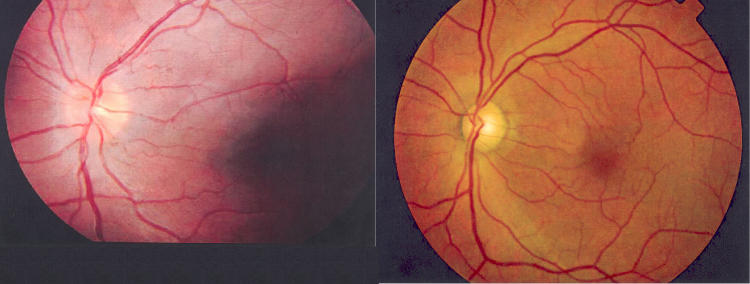
FIGURE 2.
Leber hereditary optic neuropathy. Left, Case 1. Fundus photograph taken during year 2 (2002) as part of the routine examinations of carriers. At the time, the patient had no visual complaints and visual acuities of 20/20 OU but had borderline deficiencies on FM-100 color vision testing OS. Fundus examination showed mild nerve fiber layer swelling and subtle microangiopathy both superior and inferior temporally. The patient first complained of visual loss in this eye 5 months later in 2003. Right, Case 2. Fundus photograph OS taken at year 3 (2003), about 8 months after onset of visual loss in this eye. This shows some generalized nerve fiber layer loss but relative preservation of the inferior arcuate bundle by comparison to profound papillomacular bundle loss. At this date, the patient’s visual acuity was reduced to counting fingers in this eye.

