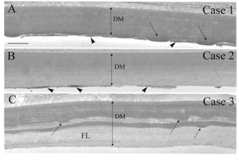
FIGURE 1.
Light microscopy of Descemet’s membrane (DM) in early-onset Fuchs corneal dystrophy (FCD). Plastic sections were stained with p-phenylenediamine to reveal compositionally distinct mixtures of extracellular matrix in DM. All three cases showed marked thickening of DM. A, Case 1 (least advanced FCD). Multiple shallow guttae (arrows) in the posterior surface, with flattened endothelial cells (arrowheads) between the guttae. Uniform staining of DM. B, Case 2 (more advanced FCD). Fewer remaining endothelial cells (arrowheads). Dark staining of anterior half of DM and slightly lighter staining in posterior half. No guttae were noted in this specimen. C, Case 3 (most advanced FCD). DM was thicker than in case 1 and case 2 and showed laminar interdigitation of dark and very lightly stained regions. Sections revealed buried guttae (arrows) covered by a lightly stained fibrillar layer (FL). All endothelial cells were missing from the posterior face of DM. Bar = 20 μm.

