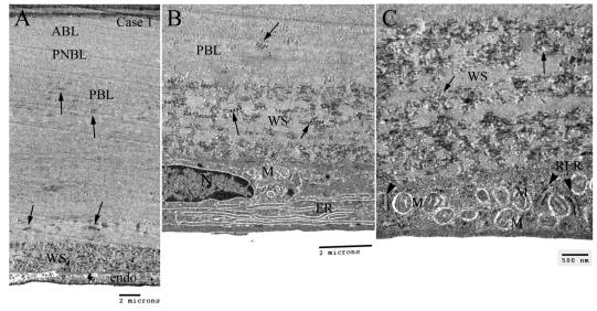
FIGURE 7.
Transmission electron microscopy showing Descemet’s membrane (DM) and endothelial cell in case 1 of early-onset Fuchs corneal dystrophy. A, Low magnification showing the full-thickness of the DM. There were many foci of wide-space collagen (arrows) in the posterior banded layer (PBL). Excessive amount of wide-space (WS) collagen were noted anterior to the endothelial cell. B, Higher magnification showing the WS collagen and an endothelial cell exhibiting abundant prominent endoplasmic reticulum (ER) and some degenerated mitochondria (M). C, Higher magnification showing the poorly formed WS collagen (arrows) and rough ER (arrowheads) and unusually abundant degenerated mitochondria (M) in the endothelial cell. ABL = anterior banded layer; PNBL = posterior nonbanded layer.

