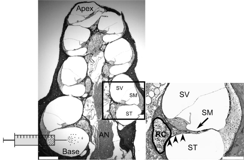
Figure 1. Anatomy of the deafened guinea pig cochlea and schematic of scala tympani cell delivery technique.

Transverse section of a guinea pig cochlea 4 weeks following ototoxic deafening, labelled with H & E. The auditory nerve (AN) is located in the middle of the structure and comprises central processes, which originate from spiral ganglion neurons (SGNs) in Rosenthal’s canal (RC; refer to inset). The AN and RC are collectively referred to as the modiolus. Encircling the modiolus are 4 cochlear turns, each comprising 3 fluid-filled compartments; scala vestibuli (SV), scala media (SM) and scala tympani (ST). The ST and SV are joined at the apex and are filled with the same fluid, perilymph. The SM is filled with endolymph, and is isolated from the ST and SV. MESCs were transplanted into the ST, at the base of the cochlea, as illustrated schematically. Scale bar = 300μm.
Inset: Higher magnification photomicrograph illustrating more comprehensively the anatomy of a single cochlear turn. The SV and SM are separated by Reissner’s membrane while the basilar membrane separates the SM and ST. Note the lack of hair cells in deafened animals (arrow), which are normally present at this location on the basilar membrane. SGNs are located within RC (circled) and are degenerating. Peripheral processes extend from SGNs toward the normal site of hair cells (arrow). RC and the ST are separated by a thin bony wall called the osseous spiral lamina (arrowheads). This wall contains numerous bony pores or canaliculae perforantes, which are thought to provide fluid communication channels between the SGNs housed within RC and the ST. Scale bar = 100μm.
