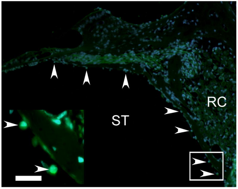
Figure 5. Survival of GFP positive MESCs in vivo.

MESCs were detected in the left cochleae of treated animals, using direct fluorescent microscopy for endogenous GFP (green) in combination with the nuclear marker DAPI (blue). Fluorescent photomicrographs illustrate MESCs in the left (treated) cochlea (arrowheads). Inset illustrates higher magnification photomicrograph of the boxed region in the image. ST = scala tympani. RC = Rosenthal’s canal. Scale bar = 20μm.
