
Figure 3. PI3K inhibition reduces pIgA transcytosis with or without Vps35 overexpression.
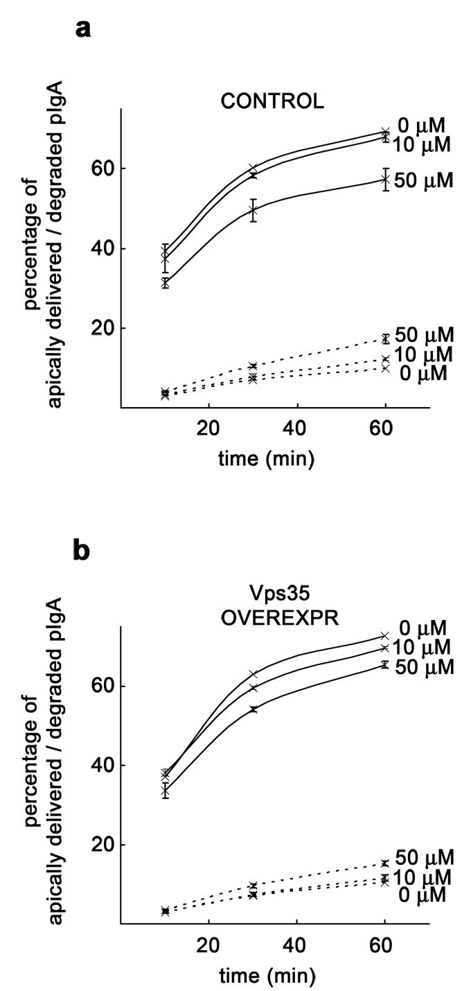
MDCK cells expressing pIgR-WT and the tetracycline transactivator (tTA) were grown as a polarized monolayer on Transwells and infected with adenovirus carrying the myc-hVps35 gene under a tetracycline repressible system. A ligand transcytosis assay using 125I-pIgA was performed. a, Decrease of apically transcytosed ligand and concomitant increase in degradation in uninduced control cells (repressed with the antibiotic) treated with 50 μM LY294002 (50 μM), with little change when a lower concentration of inhibitor (10 μM) was used. b, Decrease of apically transcytosed ligand and slight increase in degradation in cells overexpressing ~ 5-fold Vps35 (adenoviral-induced) treated with increasing concentrations of LY294002. Apical delivery is represented by a solid line and degradation by a dashed line. Values are the mean ± SD (n = 3).