
Figure 7. PI3K inhibition reduces colocalization between the two retromer subcomplexes.
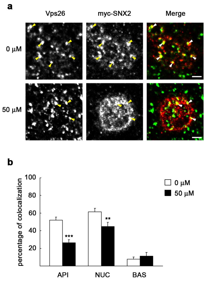
MDCK cells expressing myc-SNX2 were grown as a polarized monolayer on Transwells. After treatment with 0 μM or 50 μM LY294002, cells were fixed with paraformaldehyde and immunostained for Vps26 (green) and myc (red). a, XY sections taken at the subapical cell region show a single enlarged cell. Vps26 is distributed in this region in structures of various sizes displaying evident colocalization with myc-SNX2 (arrowheads), which was reduced upon treatment with LY294002. Scale bar represents 2.5 μm. b, The graph shows percentage of myc-SNX2 colocalizing with Vps26 throughout three representative cell levels, subapical (API), medial or nuclear level (NUC), and basolateral region (BAS); a general loss of colocalization was seen upon treatment with the inhibitor. Values are the mean ± S.E. (number of structures, n ~ 80; ** p < 0.005; *** p < 0.0005 vs. 0 μM).