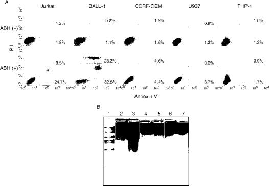
Figure 3.
ABH induced apoptosis in some cell lines. (A) Apoptosis was assessed by phosphatidylserine exposure on the cell membrane. The cells were cultured in the presence or absence of 0.1% ABH for 3 days, and stained with fluorescein isothiocyanate-conjugated annexin V and PI. The numbers in dot plots indicate the percentage of the total cell number in each quadrant. Data are representative of three separate experiments with similar results. (B) Apoptosis was also assessed by determination of DNA fragmentation. BALL-1, CCRF-CEM and THP-1 cells were cultured in the presence (lanes 3, 5 and 7) or absence (lanes 2, 4 and 6) of 0.1% ABH. Total DNA was extracted from the cells, subjected to agarose gel electrophoresis and stained with ethidium bromide. Data are representative of two separate experiments with similar results.

