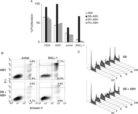
Figure 8.
p38 activation was responsible for the effects of ABH. (A) The cells were pre-treated with 10 μM SB203580 (SB), 4 μM SP600125 (SP), 10 μM PD98059 (PD) or 0.3% DMSO, which was used for dissolution of these MEK and MAPK inhibitors, for 30 min, and cultured in the presence or absence of 0.1% ABH. Tetrazolium salt was added to the cultures after 3 days. Percent proliferation indicates the proportion relative to cell growth without ABH. Data are shown as means ± SD of duplicate cultures. Data are representative of two separate experiments with similar results. (B) Jurkat and BALL-1 cells were pre-treated with SB203580 (SB) or DMSO as described above, and cultured with 0.1% ABH for 3 days. The cells were stained with fluorescein isothiocyanate-conjugated annexin V and PI. The numbers in dot plots indicate the percentage of the total cell number in each quadrant. (C) CCRF-CEM cells were enriched in S phase in advance. The cells were pre-treated with SB203580 (SB) as described above, and cultured in the presence or absence of 0.1% ABH for the indicated times (hours), followed by fixation and staining for DNA. NT indicates that the cells were not treated with thymidine and ABH. Data are representative of two separate experiments with similar results.

