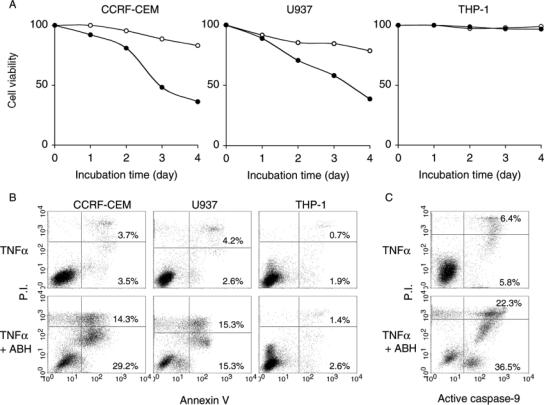
Figure 9.
The combination of ABH and TNF-α increased susceptibility to apoptosis. CCRF-CEM, U937 and THP-1 cells were cultured with 10 ng ml−1 TNF-α in the presence or absence of 0.1% ABH. (A) Cell viability was assessed by the trypan blue dye exclusion method at the indicated times (open circle, without ABH; closed circle, with ABH). Data are representative of two separate experiments with similar results. (B) The cells were stained with fluorescein isothiocyanate-conjugated annexin V and PI on day 3. (C) CCRF-CEM cells were also stained with a fluorescent-conjugated peptide specific for active caspase-9 and PI on Day 3. The numbers in dot plots indicate the percentage of the total cell number in each quadrant.

