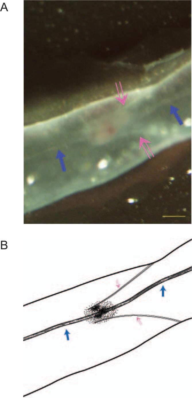
Figure 1.

A stereomicroscopic image of the lymphatic vessel around the caudal vena cava of a rat. The photograph (A) and its illustration (B) show the novel threadlike structure (solid arrow) that passes throw the lymphatic valve (open arrow). The photograph was taken in vivo and in situ, and a piece of black paper was put under the lymphatic vessel to exhibit the target clearly. The scale bar is 100 μm.
