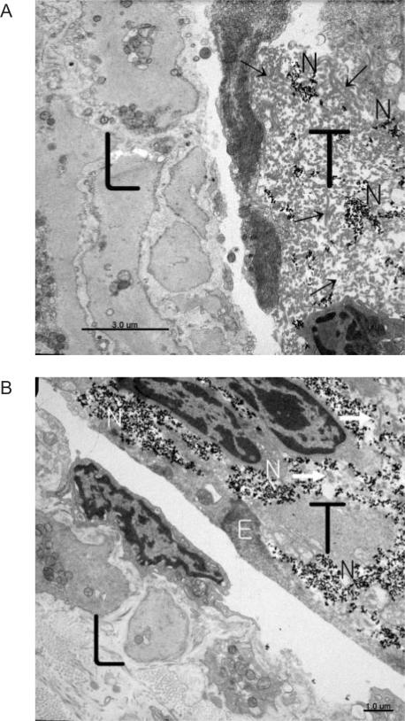
Figure 4.
TEM images of a lymphatic vessel wall and the threadlike structure which were closely located. The scattered black dots are nanoparticles. (A) On the left-hand side is the lymphatic wall in which no nanoparticles are seen. On the right-hand side is the threadlike structure in which nanoparticles (black dots) are scattered around in the exterior cellular matrix of reticular fibers. (B) Abundant nanoparticles are captured in the extracellular matrix of the threadlike structure in the upper right region. In the lower left region is the lymphatic wall where no nanoparticles are found. (Arrows are inside collagen fibrils, and L stand for lymphatic vessel; T, threadlike structure; N, nanoparticle; and E, cytoplasmic membrane of the surrounding cell of the threadlike structure.)

