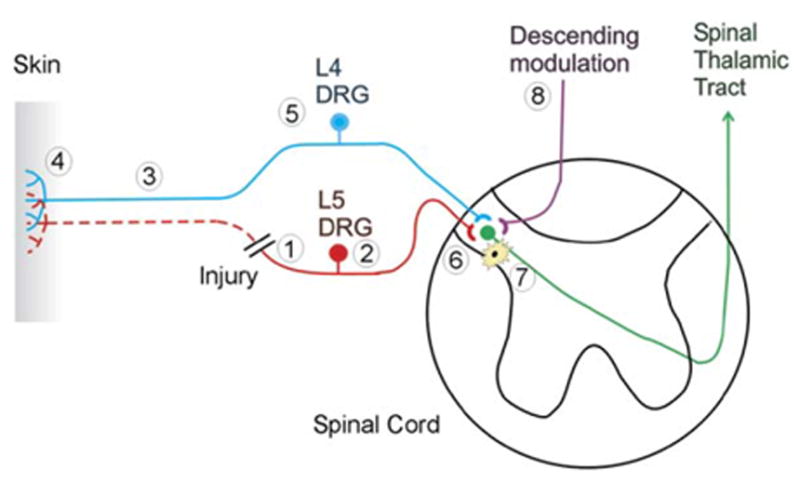
Figure 2. A Spinal Nerve Injury Leads to Alterations at Many Sites along the Neural Axis for Pain.

Eight different sites of pathophysiological changes are shown. (1) Spontaneous neural activity and ectopic sensitivity to mechanical stimuli develops at the site of nerve injury. (2) The expression of different molecules in the dorsal root ganglion of the injured nerve is up- or downregulated, reflecting the loss of trophic support from the periphery. Spontaneous neural activity develops in the dorsal root ganglia. (3) The distal part of the injured nerve undergoes Wallerian degeneration, exposing the surviving nerve fibers from uninjured portions of the nerve to a milieu of cytokines and growth factors. (4) Partial denervation of the peripheral tissues leads to an excess of trophic factors from the partly denervated tissue that can lead to sensitization of primary afferent nociceptors. (5) The expression of different molecules in the dorsal root ganglion of the uninjured nerve is up- or downregulated, reflecting the enhanced trophic support from the periphery. (6) Sensitization of the postsynaptic dorsal horn cell develops, leading to an augmentation of the response to cutaneous stimuli. (7) Activated microglial cells contribute to the development of this dorsal horn sensitization. (8) Changes in descending modulation of dorsal horn neurons also may contribute to the enhanced responsiveness of dorsal horn neurons.
