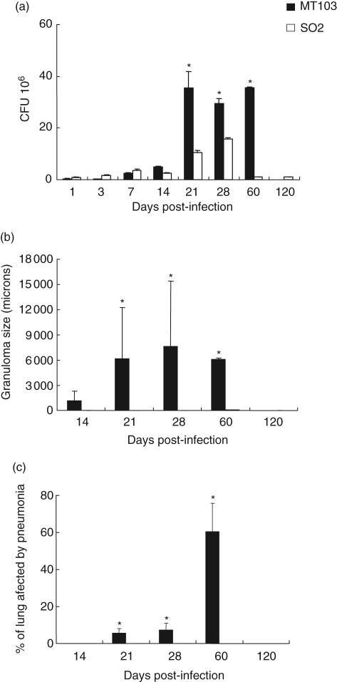
Fig. 2.
Lung bacillary burden and histomorphometry of Balb/c mice infected with Mycobacterium tuberculosis SO2 or MT103 parental strain by the intratracheal route. (a) Numbers of colony-forming units (CFUs) in the lungs of mice infected with either M. tuberculosis MT103 or M. tuberculosis SO2; (b) granuloma size (μ2) in M. tuberculosis MT103 or M. tuberculosis SO2-infected lungs; (c) percentage of lung surface area affected by pneumonia. Bars represent means ± s.d. from three mice per time-point. Asterisks represent statistical significance between groups at those time-points (P < 0·05).

