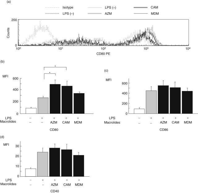
Fig. 1.
The effect of macrolides on the surface markers of co-stimulatory molecules of dendritic cells (DCs). DCs were treated without lipopolysaccharide (LPS), with LPS alone (control) or LPS with azithromycin (AZM), clarithromycin (CAM) or midecamycin (MDM). The expression of CD80 (a,b), CD86 (c) or CD40 (d) on DCs was analysed by flow cytometry after treatment. Values of mean fluorescence intensity (MFI) are expressed as the mean ± standard error of five experiments. *P < 0·05 compared with control.

