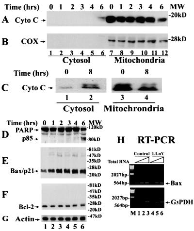
Figure 1.
Proteasome inhibitor LLnV induces Bax accumulation, cytochrome c release, and PARP cleavage in Bcl-2-overexpressing Jurkat T cells. (A–C) Jurkat T cells overexpressing Bcl-2 (0 h) were treated with 50 μM LLnV for up to 8 h, followed by preparation of cytosol and mitochondrial fractions. Both fractions were immunoblotted first by an antibody to cytochrome c (Cyto C, 17 kDa; A and C) and then reblotted by anticytochrome oxidase subunit II (COX, 26 kDa; B). Note: 20 μg protein from the cytosol, and 40 μg protein from the mitochondrial, preparation was used in each lane. (D–G) Whole-cell extracts (70 μg per lane) of the above treated cells were immunoblotted with specific antibodies to PARP (D), Bax (clone N-20; E), Bcl-2 (F), or actin (G). Molecular masses of PARP, the PARP cleavage fragment (p85), Bax, Bcl-2, and actin are 113, 85, 21, 26, and 40 kDa, respectively. Positions of protein markers are indicated at right. (H) Bcl-2-expressing Jurkat cells (Control) were treated with 50 μM LLnV for 8 h, followed by reverse transcription–PCR (RT-PCR). For the first-strand cDNA synthesis, 0.2 (lanes 1 and 4), 0.6 (lanes 2 and 5), and 1.8 μg (lanes 3 and 6) of the total RNA were used. The positions of Bax (538 bp) and glyceraldehyde-3-phosphate dehydrogenase (G3PDH) mRNA (983 bp) are indicated. Lane M is DNA molecular weight marker.

