
Fig. 5.
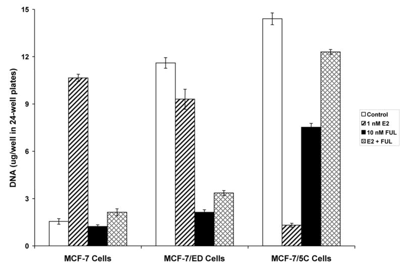
Differential proliferation of MCF-7 long-term estrogen withdrawn cell culture models in response to E2, FUL, and E2 plus FUL for 7 days. Cells were cultured under estrogen-free conditions for 4 days, and then seeded at 2 x 10⁴ cells per well in a 24-well plate. Beginning 24 hours after seeding (day 0) and every 2 days thereafter up to 6 days (days 2, 4, and 6), the cells were treated with 1 nM E2, 10 nM FUL, 1 nM E2 + 10 nM FUL, or Control (0.1% EtOH)-treated. The experiment was stopped on day 7. As a measure of proliferation, the amount of DNA per well was determined using a fluorescence-based DNA quantitation assay. Data are shown as the mean of 6 replicate wells per group ± SD. The experiment was performed 3 times independently, and one representative experiment is shown.