Figure 1.
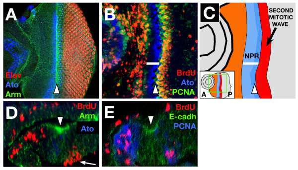
Pattern of proliferation and G1 arrest in the eye. (A) Projection of several confocal sections of a WT eye disc showing Armadillo (green), Elav (red) and Ato (blue) localization. The morphogenetic furrow (MF, arrowhead in all figures) is highlighted by accumulation of Armadillo (β-catenin), which outlines cell membranes. Ato expression appears between 6–8 cells anterior to the MF and becomes limited to the R8 photoreceptor just posterior to the furrow. Elav is expressed in all the photoreceptor cells posterior to the Ato expressing R8. In this and all subsequent figures, anterior is to the left. (B) WT eye disc showing the incorporation of BrdU (red) and the expression of PCNA-GFP reporter (indicating dE2F1 activation, green) and Ato protein (blue). Anterior to the furrow there is a non-proliferative region, without BrdU incorporation (white bar). The pattern of PCNA-GFP expression coincides with the regions of proliferation. (C) Scheme showing the different proliferating regions of the eye disc. The panel shows a drawing of the disc in 1B. The inset shows a picture of the whole disc. From anterior to posterior: orange marks the region of undifferentiated cells that proliferate randomly; the NPR is marked in blue (its extent is marked by the white bar), with the darker zone showing the morphogenetic furrow (also marked by an arrowhead); finally, the red band marks the region of the second mitotic wave. (D) Z-axis reconstruction of a WT eye disc. The outlines of the cells are shown by Armadillo (Arm) expression (green). The peripodial membrane appears at the top with some BrdU positive cells (red). In the disc proper there is a high accumulation of Arm protein in the apical part of the MF (arrowhead). S-phase nuclei in the SMW are basally located (arrow), whereas anterior to the NPR, BrdU positive nuclei are more apical. The NPR includes the furrow cells (that accumulate high apical levels of Arm) and between 4–6 rows of more anterior cells. Ato expressing cells (blue) are restricted to the disc proper. (E) The cytoplasmic expression of PCNA-GFP reporter (blue) is seen in cells that are completing the cell cycle in the most anterior part of the NPR, but is not expressed in the cells of the MF (that accumulate E-cadherin in the apical region, in green). In the SMW the cells with BrdU positive nuclei also express the PCNA-GFP.

