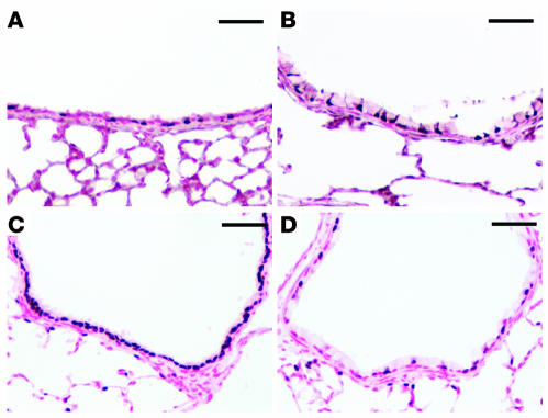
Figure 9. FOXJ1 and loss of FOXA2 staining in lungs of CCSP-rtTA/TRE2-Spdef transgenic mice.
Immunohistochemistry for FOXJ1 (A and B) and FOXA2 (C and D) was performed on the lung sections of control mice (A and C) and transgenic mice expressing SPDEF (B and D). The normal staining pattern of FOXJ1, a ciliated cell marker, was unaltered by expression of SPDEF (A and B). FOXA2 staining was observed throughout the epithelium in control mice (C). FOXA2 was not detected in the goblet cells lining the conducting airways of the transgenic mice, but persisted in ciliated cells (D). Scale bars: 50 μm.

