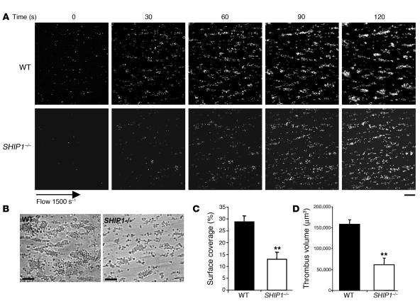
Figure 2. Thrombus formation defect in SHIP1-deficient platelets under arterial flow conditions.
DiOC6-labeled platelets in whole blood were perfused through a collagen-coated microcapillary at a shear rate of 1,500 s–1 during 2 minutes. (A) Thrombus formation was visualized with a ×40 long-working-distance objective in real time and then imaged by transmitted light microscopy (B) (scale bar: 20 μm). (C and D) Area covered by platelet thrombi (C) and thrombus volume (D) were measured at 2 surface locations in each of 3 different experiments (mean ± SEM). **Significant difference (P < 0.005) versus WT, according to 1-tailed Student’s t test.

