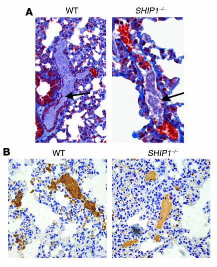
Figure 6. Histology and immunohistochemistry of WT and SHIP1-deficient mice injected with collagen and epinephrine.
(A) Sections of lung tissue were stained with Masson trichrome. Arrows point to thrombi in the pulmonary vasculature of WT and SHIP1-deficient mice. Original magnification, ×200. (B) Platelet staining (brown) revealing extensive platelet thromboemboli in WT mice and a decrease in platelet number in thrombi formed in SHIP1-deficient platelets. Original magnification, ×200.

