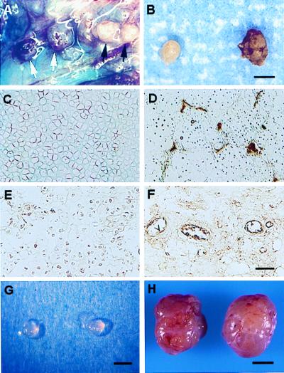
Figure 1.
Distinctive avascular and vascular phenotypes of chondrosarcoma tumor nodules. (A) Injection of tumor cell suspensions into air sacs in the sacral region of rats resulted in the formation of distinct tumor nodules, which could be visually distinguished from each other on the basis of their degree of vascularization i.e., vascular (white arrows) from avascular (black arrows). At day 10, approximately 50% of the tumor nodules were vascularized and by day 12, the majority of the nodules were vascularized. These results were consistently observed throughout more than 15 independent experiments. (B) Representative avascular and vascular nodules, respectively, are shown. Factor VIII immunostaining demonstrates the avascularity of representative control xiphoid cartilage sections (C) in which microvessels are not detected, in contrast to sections of chondrosarcoma in which a number of microvessels are easily seen (D). A lack of microvessels is observed in avascular chondrosarcoma nodules (E) when compared with vascular nodules in which positive microvessel staining is apparent (F). When avascular tumor nodules (G) were dissected from air sacs and implanted s.c. in rats, the majority of avascular nodules (11/13) became vascularized tumors (H). (Scale bars: A and B, 2 mm; C–F, 100 μm; G, 2 mm; H, 10 mm.)

