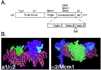
Figure 1.
α2 contains a surface exposed PTS1-like sequence in the homeodomain. (A) A schematic diagram of α2 illustrating its domain structure. Interacting proteins are placed above the domain where they are known to make contacts. The homeodomain is shown in more detail in the Inset. The PTS1-like sequence is placed below its position in the linear sequence of the homeodomain. (B) The PTS1-like sequence in the homeodomain of α2 is surface exposed. (Left) The structure of the α2 homeodomain (purple) is shown bound as a trimeric complex with the homeodomain of a1 (green) and DNA (brown) (21). (Right) The α2 homeodomain (purple) is shown bound to DNA (brown) and a dimer of Mcm1 (green) (20). In each of the structures the PTS1 sequence in the homeodomain of α2 is shown in blue.

