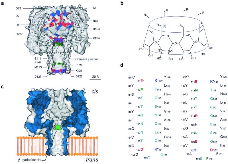
Figure 1.
Representations of the proteins and cyclodextrins used in this work. (a), Sagittal section through the wt-αHL pore showing the location of all of the charged side chains in the channel lumen (red, negative; blue, positive) and the key hydrophobic residues M113 and L135 (green). The site of the junction in the chimera αHL-CH1 is indicated; (b), Structures of the βCDs used in this work. βCD, R = −OH; s7βCD, R = −OSO3−; (c), Schematic of the wt-αHL pore showing βCD lodged in the lumen of the channel. The location is based on mutagenesis data (16); (d), Sequences of the transmembrane β barrels in wt-αHL (Left) and αHL-CH1 (Right).

