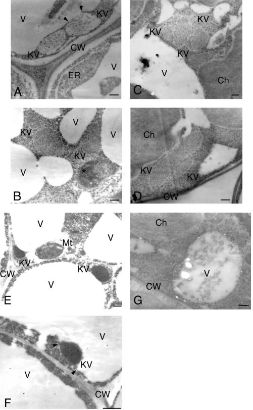
Figure 2.
Electron micrographs showing development of KV-like vesicles in cells of several tissues from transgenic Arabidopsis expressing wild-type SH-EP. A, SH-EP accumulated in ER and KV-like vesicles in stem cells. A KV-like vesicle fused with a vacuole (arrowheads). B, SH-EP was accumulated in KV-like vesicles in cotyledon cells. C, SH-EP was accumulated in KV-like vesicles in rosette leaf cells. D, Enlarged and oblong-shaped KV-like vesicles were observed in rosette leaf cells. E, SH-EP accumulated in KV-like vesicles in sepal cells. F, A KV-like vesicle was fused with vacuoles in sepal cells. Arrowheads indicate possible fusion sites. G, SH-EP was also localized in vacuoles of cells of rosette leaves. Ch, Chloroplast; CW, cell wall; KV, KV-like vesicle; Mt, Mitochondrion; V, vacuole; An asterisk indicates an unidentified cell compartment. Bars = 200 nm.

