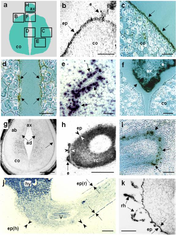
Figure 5.
Transcript distribution of VfPTR1 in sections of developing faba bean seeds and seedlings. a, Schematic representation of a developing faba bean embryo in a cross-section, where the boxed areas are enlarged in the following pictures; b through k, Bright-field micrographs showing in situ hybridization using a 33P-labeled VfPTR1 probe, with label seen as dark grains; b, abaxial region of a stage IV (Borisjuk et al., 1995) cotyledon with labeled epidermal cells and endosperm, but unlabeled parenchyma cells; c and d, abaxial and adaxial regions of cotyledons, respectively. The abaxial transfer cells in the outer epidermis are more strongly labeled than the cells of the inner epidermis; e, labeled cells surrounding transport vessels of a stage VI cotyledon; f, strong labeling of endosperm surrounding the axis during stage VI; g, cotyledons with labeled storage parenchyma cells and transfer cells of the outer epidermal region; h, radicle of a stage VII embryo strongly labeled within the cortex cells, but not the epidermis cells (seen as an unlabeled layer between the cortex and the endosperm). i through k, Distribution of VfPTR1 transcripts in sections of faba bean seedlings; i, section of a root showing the labeled vascular bundles; j, longitudinal section through the seedling, showing strong label in the hypocotyl and in the epidermis of the root, but not in the epidermis of the hypocotyl; k, cross-section through the root of a seedling showing labeling within the epidermis and the root hairs; ab, Abaxial region; ad, adaxial region; ax, axis; co, cotyledons; e, endosperm; ep, epidermis; ep(h), epidermis of hypocotyl; ep(r), epidermis of root; hy, hypocotyl; rc, radicle cortex; rh, root hair; v, vascular bundle. Bars in b, c, d, e, f, i, and k = 0.1 mm; g and j = 1 mm; h = 0.5 mm. Arrows point to the labeling and arrowheads point to unlabeled areas.

