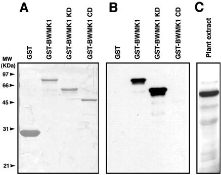
Figure 3.
Specificity of Ab-pNBWMK1 antibody. A, SDS-PAGE analysis of GST fusion recombinant proteins of BWMK1. Purified proteins were separated on a 10% (w/v) SDS-PAGE and stained with Coomassie Brilliant Blue R-250. The apparent molecular masses (kilodaltons) are indicated at the left. B, Immunoblot analysis using the recombinant protein. Ab-pNBWMK1 specially recognized GST-BWMK1 and GST-BWMK1 KD. C, Immunoblot analysis using plant extracts from rice. Protein (50 μg) was separated by SDS-PAGE, blotted, and probed with Ab-pNBWMK1 (1:3,000 [v/v] dilution). After incubation with a horseradish peroxidase-conjugated secondary antibody, the complex was visualized using enhanced chemiluminescence.

