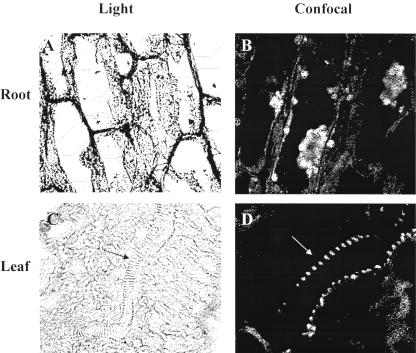
Figure 2.
Immunohistochemical studies profiled MIPS expression in organs of bean. Sections of 8-d-old bean roots (A and B) and leaves (C and D) were stained with toluidine blue and incubated with MIPS antibody and fluorescein isothiocyanate-conjugated secondary antibody. Shown are light (A) and confocal (B) micrographs of a longitudinal section of root and light (C) and confocal (D) micrographs of leaf section (arrows show vascular system). Root and leaf light micrographs were magnified 580× and 370×, respectively. Bar = 20 μm. Magnification of confocal micrographs could not be determined with the Nikon PCM 2000 microscope. Controls included unstained sections not treated with primary or secondary antibody to detect autofluorescence and stained sections incubated only with secondary antibody to detect nonspecific interactions.

