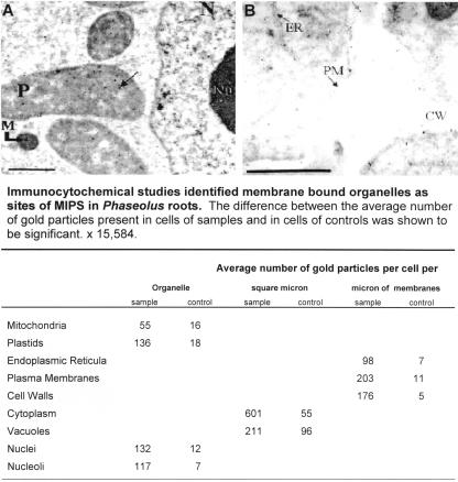
Figure 3.
Immunocytochemical analyses identified membrane-bound organelles as subcellular locations of MIPS. Bean root sections were incubated with MIPS primary antibody and goat anti-rabbit 10-nm gold-conjugated secondary antibody and photographed with a Zeiss 10A TEM microscope. Controls were incubated with goat anti-rabbit 10-nm gold-conjugated secondary antibody only. As shown in A and B, gold particles are present in plastids (P), mitochondria (M), the nucleus (N), nucleolus (Nu), endoplasmic reticula (ER), plasma membrane (PM), and the cell wall (CW). Bar = 1 μm.

