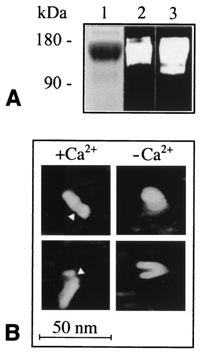
Figure 1.
Purified VE-cadherin-Fc characterized by SDS/PAGE and immunoblotting (A) and by AFM imaging (B). (A) SDS/PAGE was stained with Coomassie blue (1), and immunoblots were probed with VE-cadherin antibody (mAb 11D4.1) (2) and antibody against human IgG (3). Electrophoretic mobility (160–180 kDa) corresponds exactly to the calculated molecular weight of the chimeric dimer. (B) AFM images of hydrated VE-cadherin-Fc adsorbed to mica and scanned in the presence and absence of Ca2+ in isotonic buffer. Note elongated rod-like structure in Ca2+ and globular to v-shaped morphology in the absence of Ca2+. Arrowheads point to constrictions assumed to mark the boundary between the Fc-portion and the cadherin moiety.

