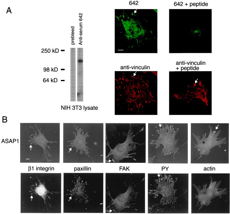
Figure 1.
ASAP1 associates with FAs. Arrows point to FAs. (A) Antibody specific for ASAP1. Antiserum 642 was used for Western blotting (Left) at a 1:10,000 dilution and for immunofluorescence (Right) at a dilution of 1:2,000. Cell lysate (1 μg) was used for Western blotting. The peptide (500 μM) to which the antibody was raised was included where indicated. (B) Colocalization of ASAP1 with markers of FAs. PY, protein phosphotyrosine. (Bars = 10 μm.)

