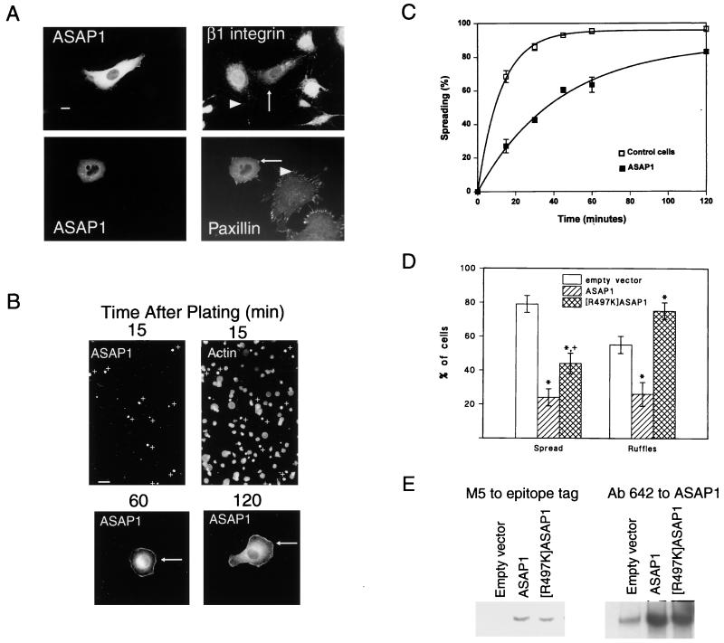
Figure 3.
Overexpression of ASAP1 affects cytoskeletal remodeling. NIH 3T3 fibroblasts were transfected with plasmids for FLAG-tagged ASAP1. (A) FA morphology. For double staining for β1 integrin, ASAP1 was detected by using a monoclonal antibody to the FLAG epitope. For paxillin, ASAP1 was detected with the polyclonal antibody 642. In this case, endogenous ASAP1 was apparent in FAs in nontransfected cells, visible in photographs in which the transfected cells were overexposed. (Bar = 10 μm.) (B) Comparison of transfected and nontransfected cells during spreading. A low-power field photographed from experiments used to derive the data for C is shown. (Bar = 40 μm.) At higher magnification, two transfected cells, fixed 1 and 2 h after plating, are shown. (C) Effect of ASAP1 overexpression on cell spreading rate. NIH 3T3 fibroblasts were seeded on fibronectin-coated coverslips. At the indicated times, cells were removed and fixed. Transfected cells were detected by using a monoclonal antibody to the FLAG epitope and were counterstained with rhodamine-phalloidin. A cell was considered spread when the total diameter was 2-fold or greater than the nuclear diameter. The data are the means and range for two experiments. (D) Effect of ASAP1 and [R497K]ASAP1 overexpression on cell spreading and PDGF-induced dorsal ruffles. Cells were transfected with plasmids encoding FLAG epitope-tagged ASAP1 or [R497K]ASAP1. Spreading was determined at 15 min. To examine ruffling, cells were treated for 4 min with 10 ng/ml PDGF. Dorsal ruffles were visualized by using rhodamine-phalloidin. Data are shown as mean ± SD for four experiments. ∗, Different from control, P < 0.001; +, different from wild type, P < 0.01. (E) Expression levels of FLAG-tagged ASAP1 and FLAG-tagged [R497K]ASAP1. Cells were transfected in parallel to those used for spreading and ruffling assay and lysed in RIPA buffer (0.15 mM NaCl/0.05 mM Tris⋅HCl, pH 7.2/1% Triton X-100/1% sodium deoxycholate/0.1% SDS). ASAP1 in 1 μg of cell lysate protein was detected with an antibody to the FLAG epitope (Left) and in 0.1 μg of cell lysate protein with antibody 642 (Right).

