Figure 1.
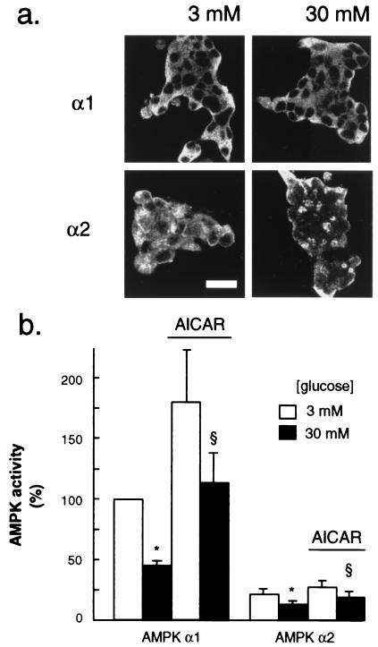
Immunolocalization of AMPK isoforms in MIN6 beta cells (a) and regulation of AMPK activity by glucose and AICAR (b). (a) Cells were incubated for 24 h at the indicated glucose concentrations before probing for AMPK α1 and α2 isoforms as detailed in Experimental Procedures. Shown are clusters of 30–40 cells; the dark areas in the center of each cell, most evident after probing with the anti-AMPK α1 antibody, correspond to the position of the nuclei, as identified in bright-field images (not shown). (Bar = 20 μm.) (b) After incubation for 6 h at the indicated glucose concentration, each isoform of AMPK activity was assayed after immunoprecipitation as given in Experimental Procedures. Data are normalized to activity vs. AMPK α1 in extracts cultured at 3 mM and represent the means ± SEM of four separate experiments. P < 0.05 for the effect of 30 mM glucose (*) and 200 μM AICAR (§).

