Figure 1.
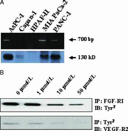
Expression of FGF-RI in pancreatic cancer cell lines. (A) mRNA was isolated, and RT-PCR was performed. After PCR with specific primers for FGF-RI, the PCR product was separated on a 1% agarose gel (upper panel). The corresponding FGF-RI Western blot analysis is shown below (lower panel). In (B), the effect of PD173074 on FGF-RI and VEGF-RII signaling is shown. AsPC-1 cells were grown for 24 hours in the presence or in the absence of PD173074 and were stimulated for 15 minutes with FGF-1 (50 µg/ml) in the presence of heparin or stimulated with recombinant human VEGF (25 ng/ml). Total cell lysates were immunoprecipitated (IP); subsequently, immunoblotting (IB) was performed with the indicated antibodies.

