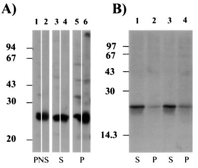
Figure 1.
Distribution of GAIP in AtT-20#1 cells. (A) AtT-20 cells stably expressing GAIP (clone #1) were homogenized, and the postnuclear supernatant (PNS) was centrifuged at 100,000 × g to yield crude membrane (P) and cytosolic fractions (S). Then, 25 μg of protein/lane were separated by SDS/PAGE, cut in two strips, and immunoblotted with anti-GAIP (N) (lanes 1, 3, and 5) or anti-HA (lanes 2, 4, and 6) and detected by enhanced chemiluminescence. GAIP is found in the PNS (lanes 1 and 2) and in both cytosolic (lanes 3 and 4) and membrane (lanes 5 and 6) fractions. (B) Cells were biosynthetically labeled for 4 h with Easytag, homogenized and fractionated as in A, and solubilized in RIPA buffer. GAIP was immunoprecipitated from the cytosolic (lanes 1 and 3) or crude membrane (lanes 2 and 4) fraction with anti-GAIP (23-217) (lanes 1 and 2) or anti-GAIP (C) (lanes 2 and 3). Immune complexes were separated by SDS/PAGE and detected by autoradiography. Newly synthesized, [35S]GAIP was found mainly in the cytosolic fraction.

