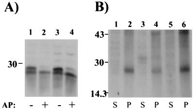
Figure 2.
The membrane pool of GAIP is phosphorylated. (A) A total of 25 μg (lanes 1 and 2) or 50 μg (lanes 3 and 4) protein from the RM fraction of rat liver were incubated for 3 h with 30 units alkaline phosphatase (AP), separated by SDS/PAGE, followed by immunoblotting with anti-GAIP (N) and enhanced chemiluminescence. After alkaline phosphatase digestion, the slower migrating band (26.5 kDa) of GAIP disappears and only the faster migrating band (28.5 kDa) is seen. (B) AtT-20#1 cells were labeled in vivo with [32P]orthophosphate for 4 h, and cytosolic (lanes 1, 3, and 5) and membrane fractions (lanes 2, 4, and 6) were prepared and solubilized as in Fig. 1. GAIP was immunoprecipitated using anti-HA (lanes 1 and 2), anti-GAIP (23–217) (lanes 3 and 4), or anti-GAIP (N) (lanes 5 and 6). Immune complexes were resolved by SDS/PAGE and exposed overnight for autoradiography. Phosphorylated GAIP is detected exclusively in the membrane fraction (P) with all three antibodies.

