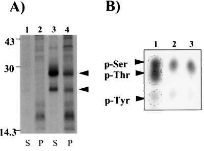
Figure 3.
Immunoprecipitation and phosphoamino acid analysis of 32P-labeled GAIP in transiently transfected HEK 293T cells. (A) Cells were transiently transfected with a mock pcDNA3 (lanes 1 and 2) or with pcDNA3 GAIP (lanes 3 and 4) and labeled with [32P]orthophosphate for 2 h. GAIP was immunoprecipitated from cytosolic (S) and membrane (P) fractions with anti-GAIP (C) as in Fig. 1. 32P-labeled GAIP (arrows, lanes 3 and 4) is found in both cytosolic (S) and membrane (P) fractions. A faster moving band, 23.1 kDa, is immunoprecipitated from cells labeled with [32P]orthophosphate after transfection with GAIP (28 kDa), probably resulting from an alternative initiation start site (25). (B) In the last 15 min of labeling, cells were incubated with buffer as control (lane 2) or 150 nM PMA (lane 3). The 32P-labeled GAIP bands were cut out from both the cytosolic and membrane fractions and used for one-dimensional phosphoamino acid analysis on a cellulose-coated plate as described in Experimental Procedures. Lane 1: p-Ser, p-Thr, and p-Tyr standards detected by ninhydrin staining (arrowheads, lane 1). Lanes 2 and 3: Autoradiography p-Ser was the major phosphoamino acid present in both control (lane 2) and PMA-treated cells (lane 3).

