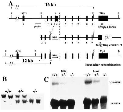
Figure 1.
Targeted inactivation of the MT1-MMP gene. (A) 5′ region of the wild type (Top), targeting construct (Middle), and the gene region after the targeting event. Exons are shown as black boxes and numbered. Directions of the neo and tk genes (open boxes) are depicted by arrows. The probe used for Southern blots and the location of primers used for PCR (short solid lines) are indicated. E, EcoRI; S, StuI; B, BamHI. (B) Southern blot analysis of EcoRI-digested DNA from a litter generated by F1 heterozygote mating. The wild-type fragment is 16 kb, and the mutant one is about 12 kb. (C) Northern blot analyses of RNA isolated from wild-type and null mouse lung and heart tissues. The filter was hybridized with an MT1-MMP cDNA probe, a probe for 18S RNA being used as a control for RNA loading and integrity.

