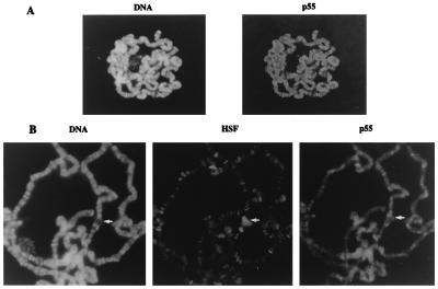
Figure 4.
Immunofluorescent localization of p55 on polytene chromosomes. (A) Indirect immunofluorescent staining for p55 in a polytene nucleus (Left) and DNA staining (Right). Antibody specificity was demonstrated by the inhibition of immunoreactivity by the inclusion of purified recombinant p55 protein with anti-p55. (B) Indirect immunofluorescent staining for p55, HSF, and DNA on polytene chromosomes prepared from heat-shocked third instar larvae. The heat shock puff at 87C is indicated by the arrow. We have noticed several instances where p55 appears more localized to a few specific sites above the general nonspecific staining; the reproducibility and cytological location of this staining require further study for clarification.

