Fig. 3.
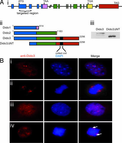
Targeted Dido gene disruption results in an N-terminal-truncated Dido3 form that loses centrosome association. (Ai) Dido gene architecture. A common N-terminal domain (blue) is followed by a specific C-terminal domain for each of the three splice variants (pink, green, yellow, and red). The domain targeted in mutant mice is indicated. (Aii) Dido3ΔNT is the truncated Dido3 form in mutant cells after disrupting the targeted region of the Dido gene, as in Ai. (Aiii) Western blot of WT and Dido-targeted (mutant) MEF lysates stained with anti-Dido3. The lower band is the truncated form of Dido3. (B) MEFs were stained with anti-Dido3 (red) and DAPI (blue). (Bi and Bii) In interphase (Bi) and mitotic (Bii) WT cells, Dido3 clearly associates with centrosomes/spindle poles. (Biii) In interphase mutant cells, the Dido3-truncated form shows a diffuse nuclear localization without the two-dot centrosome pattern. (Biv) In mitotic mutant cells, there is no spindle pole association and chromosomes appear to be misaligned (arrows).

