Fig. 4.
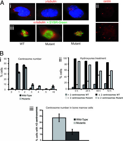
Targeted Dido disruption leads to centrosome amplification and spindle malformation. (Ai) Anti-γ-tubulin staining revealed mutant MEFs with 4, 8, or 12 centrosomes. (Aii) Anti-centrin staining confirmed centrosome amplification in mutant cells (Lower), whereas WT MEFs showed normal numbers (Upper). (Aiii) MEFs were stained with anti-α-tubulin (red) to show spindle formation. DNA was stained with SYBR-Green. WT MEFs (Left) had normal spindle formation. Mutants formed a spindle with four poles (Center) or a completely disorganized spindle (Right). (Bi) We counted centrosomes in anti-γ-tubulin-stained WT and mutant MEFs. The x axis shows centrosomes per cell. Approximately 23% of Dido-mutant cells had three or more centrosomes, whereas only 3.9% of WT cells showed this abnormal number. (Bii) Percentage of WT and mutant MEFs showing ≤2 or >2 centrosomes per cell after hydroxyurea treatment. The percentage of mutant cells with aberrant centrosome numbers increased with time, reaching >40% of total cells at 48 h. The x axis depicts time of hydroxyurea treatment. (Biii) Freshly isolated BM samples stained with anti-γ-tubulin. A significantly larger percentage of cells from mutant mice had more than two centrosomes compared with cells from WT mice (Student's t test, P = 0.008).

