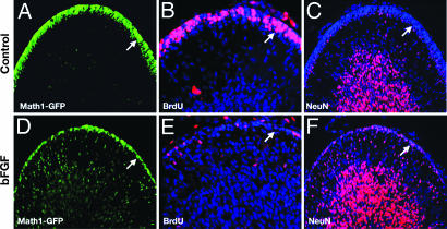
Fig. 6.
bFGF accelerates differentiation of GCPs in vivo. Math1-GFP mice were given intracisternal injections of vehicle (Control, A–C) or bFGF (D–F) on P4, P5, and P6, and i.p. injections of BrdU on P6. At P7, cerebella were fixed and stained with anti-GFP (A and D), anti-BrdU (B and E), or anti-NeuN antibodies (C and F) with a DAPI counterstain (blue). Note the reduced number of immature, proliferating (GFP-high, BrdU+) cells and the abundance of differentiated (NeuN+) cells in the EGL of FGF-treated mice (see arrows in A vs. D, B vs. E, and C vs. F). Data are representative of six mice.

