Fig. 5.
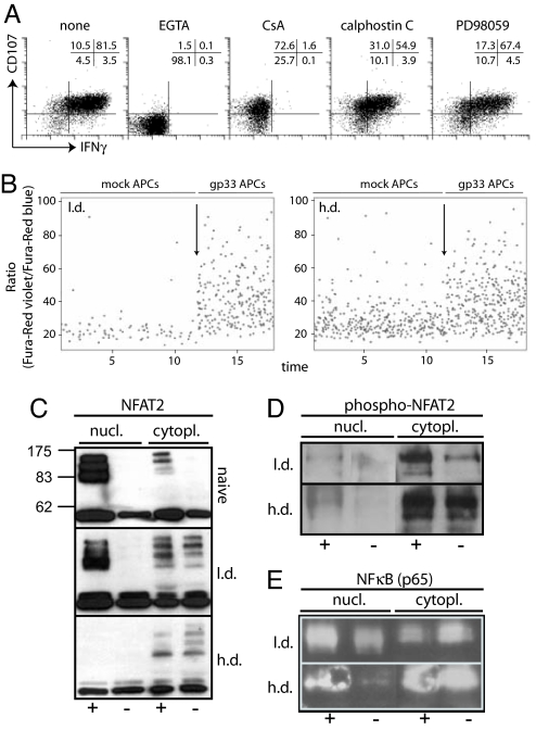
TCR signaling in LCMV-specific CD8+ T cells from h.d. or l.d. infected mice. (A) Signaling requirements for degranulation and IFN-γ production. Spleen cells from adoptively transfused and infected mice (day 20) were stimulated with gp33 peptide in the presence of EGTA, cyclosporine A, calphostin C, or PD98059 and degranulation and IFN-γ production were analyzed. Plots are gated on Ly5.1+ TCR tg CD8+ T cells. One representative staining of two mice and three independent experiments is shown. (B) Ca2+ flux analysis. Spleen cells were isolated, labeled with Fura-red, and stained for Ly5.1 followed by incubation with mock- or gp33-pulsed C57BL/6 peritoneal macrophages; arrows indicate the start of analysis of Ca2+ flux in the presence of gp33-pulsed macrophages. Plots are gated on Ly5.1+ T cells. Representative plots of two mice and three independent experiments are shown. (C–E) Ly5.1 TCR tg CD8+ T cells were sorted from adoptively transfused and h.d.- or l.d.-infected mice or from naïve TCR tg mice. Sorted cells were incubated in presence (+) or absence (−) of gp33 peptide for 16 h. Nuclear and cytoplasmic extracts were prepared and assayed in Western blots for presence of NFAT2 (C; molecular mass, 90, 110, or 140 kDa), p-NFAT2 (D), or NF-κB (E). One of two or three similar experiments is shown.

