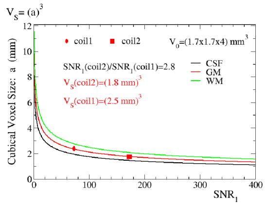
Figure 2.

A plot of the suggested cubical voxel size versus SNR0 for three different brain tissues (Equation [5] simulation) is shown. The imaging voxel volume V0 matches the volume used in experiments, and the TSNRL or 1/λ values obtained in experiments were used (GM: 80, WM 130, CSF 45). The suggested voxel volumes for two different coils: a standard system provided birdcage head coil- coil1 (birdcage) and a 16 channel receive-only surface coil brain array- coil2 (array) are shown in gray matter (coil1 marked as a red oval, coil2 marked as a red square). Coil2 (array) has 2.8 times better SNR over the whole brain than coil1 (birdcage).
