
Figure 4.
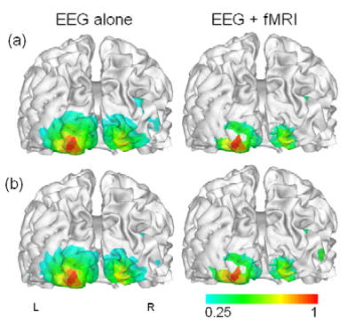
Estimated cortical source images from Subject VG’s VEP: (a) recorded outside MRI scanner; (b) recorded inside MRI scanner during fMRI scanning. The contour scales are normalized with respect to the maximum value.
Official websites use .gov
A
.gov website belongs to an official
government organization in the United States.
Secure .gov websites use HTTPS
A lock (
) or https:// means you've safely
connected to the .gov website. Share sensitive
information only on official, secure websites.
Estimated cortical source images from Subject VG’s VEP: (a) recorded outside MRI scanner; (b) recorded inside MRI scanner during fMRI scanning. The contour scales are normalized with respect to the maximum value.