Figure 8.
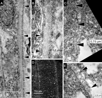
Ultrastructure of the notochord sheath reveals patterns of stress that correlate with rings of phosphorylated Fak. (A) TEM of a horizontal section through the notochord of a prim-5 embryo (24 hpf). Black arrowheads indicate changes in the orientation of fibers in the inner layer. The notochord is to the left of the sheath and somites to the right. (B) High-magnification TEM of this notochord sheath showing sites where the membrane of a notochord cell adheres to the fibrous sheath (black arrowheads). (D) Projection of confocal sections of the notochord of a prim-5 embryo stained with anti-pY861Fak shows circumferential striations (anterior up, dorsal to the right). (C) TEM of a horizontal section through the notochord of a three-somite embryo (11 hpf) showing the immature notochord sheath marked with black arrowheads. The notochord cells are to the left of the sheath. (E) High-magnification TEMs of the notochord sheath (black arrowheads) in a three-somite embryo showing only circumferential fiber orientations.

