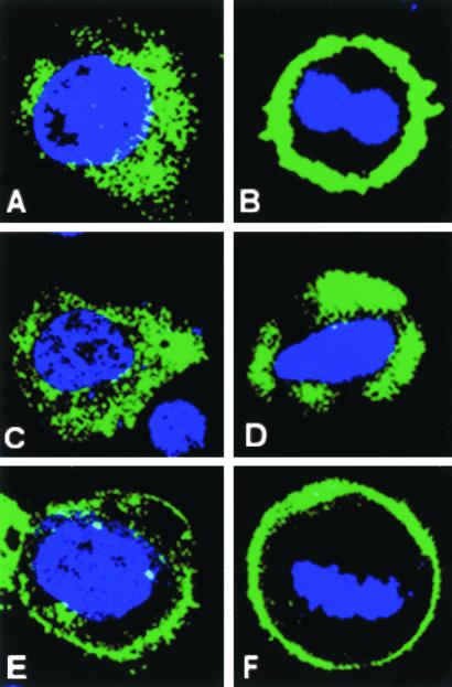
Figure 2.
Domain dissection of LGN. FLAG-tagged LGN constructs were transfected into WISH cells, and their subcellular localization was detected using anti-FLAG antibody (green). (A) Perinuclear/cytoplasmic staining for FL-LGN at interphase; (B) a cortical localization for FL-LGN at metaphase. N-FLAG (amino acids 1–384) remains mainly in the cytoplasm during interphase (C) and metaphase (D). C-FLAG (amino acids 385–672) is enriched at the cell cortex during both interphase (E) and metaphase (F). Residual cytoplasmic/perinuclear staining for C-FLAG can also be seen at interphase (E). The cell cycle stage was determined by DNA staining (blue).

