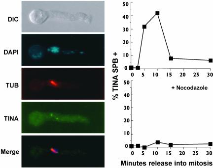
Figure 4.
Micrograph shows that TINA localizes to the ends of spindles at mitosis. A metaphase cell is shown with, from the top, a differential interference contrast image, DAPI staining revealing condensed DNA, tubulin staining of the mitotic spindle, TINA staining, and a merge. The graph at right shows the kinetics of localization of TINA to spindle poles during synchronous mitosis. A strain containing the nimT23 mutation that expressed HA-tagged TINA was arrested at G2 by shift to 42°C for 3 h (time 0) before down-shift to 30°C to allow entry into mitosis (as in Figure 3). The percentage of cells displaying SPB localization of TINA was determined using immunofluorescence. Another culture, as indicated, was treated with nocodazole before release into mitosis.

