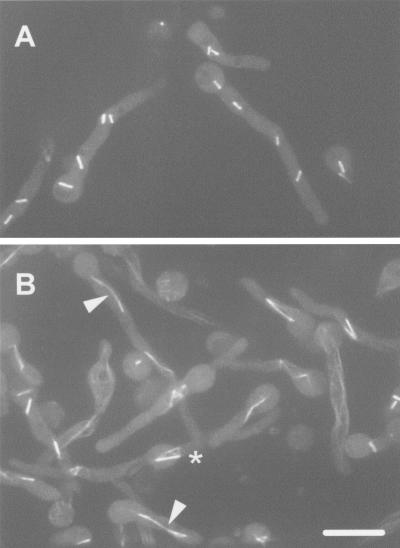
Figure 7.
Live cell microtubule architecture during a bimE7 imposed metaphase arrest with tinA (A) and without tinA function (B). Cells containing bimE7 + GFP-tubA (A) or bimE7/ΔtinA + GFP-tubA (B) were shifted to 42°C to impose metaphase arrest by inactivation of bimE7. Cells were viewed using a spinning disk confocal microscope and the micrographs are maximum intensity projections of a Z-series stack taken from video-C.mov (A) and video-D.mov (B) from the 15-min exposure (910 s) during recordings >40 min. Arrowheads in B indicate spindle joined by astral microtubules. The * indicates parallel spindles with their astral microtubules interacting which form a square like structure by the end of the video. Bar, ∼5 μM.

