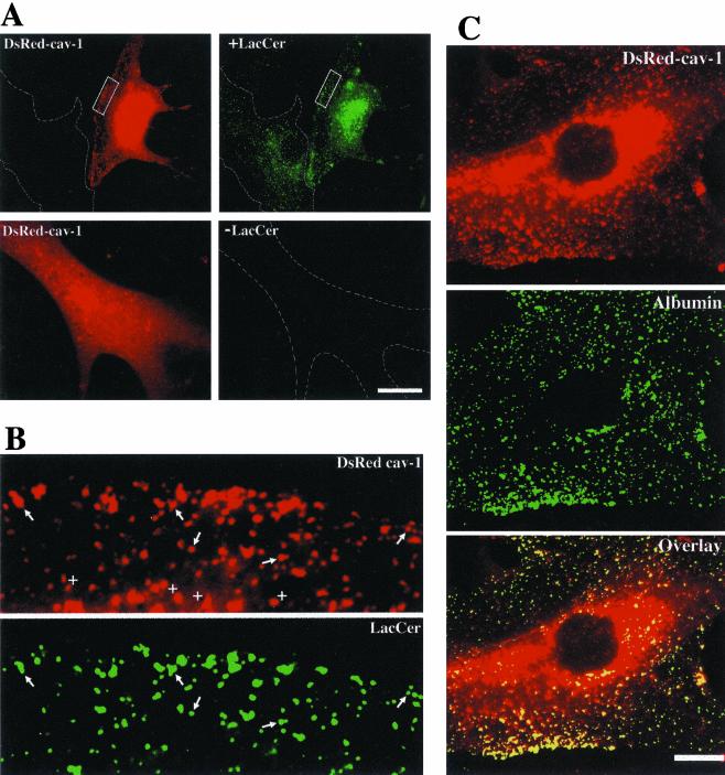
Figure 4.
DsRed-cav-1 colocalization with BODIPY-LacCer and fluorescent albumin. RFs were transfected with DsRed-cav-1 and 48 h later the distribution of (A and B) BODIPY-LacCer or (C) AF488 albumin after 2 min of internalization at 37°C was examined. Top panels in A show the same field for cav-1 (red) or LacCer (green). Note that in an adjacent nontransfected cell outlined in white, there was no spillover of LacCer fluorescence into the DsRed channel. Bottom panels in (A) show a control experiment using transfected cells without LacCer labeling. Note that no DsRed fluorescence appeared in the green channel at the exposure setting used for LacCer in the doubly labeled specimen. The boxed regions in A are shown at higher magnification in B. Note the similar patterns of punctate fluorescence (e.g., at arrows) for LacCer and cav-1. Quantitative analysis indicated that at least ∼90% of the green puncta colocalized with DsRed-cav-1. However, not all DsRed-cav-1 puncta were positive for LacCer (e.g., see +s). (C) Fluorescence micrographs of a cell expressing DsRed-cav-1 and labeled with AF488 albumin. More than 95% of the AF488 puncta colocalized with DsRed-cav-1. Bars, 10 μm.

